


Clinical Cognitive Neuroscience Center
Research
Neuromodulation
Neuromodulation
Key question. How do brain stimulation and neurosurgical treatments change brain circuits, and can these changes tell us who will respond and where to stimulate?
Approach. We study patients with schizophrenia spectrum disorder with MRI and EEG before and after electroconvulsive therapy (ECT), comparing them with patients treated with medication alone. We also study rTMS and tDCS, and Gamma Knife surgery and deep brain stimulation for treatment-refractory OCD. Building on ECT-induced circuit changes, we are developing personalized stimulation targets using individualized head models.
Recent Findings
Treatment-Related Changes and Neuromodulation Targets
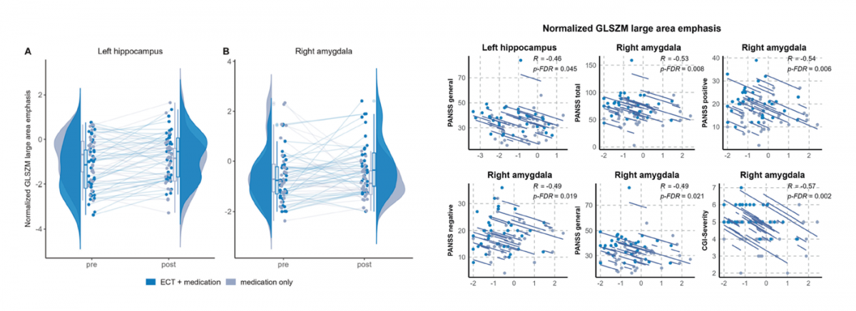
In patients with psychosis, MRI texture changes in limbic gray matter after ECT were associated with clinical response (Choe et al., Mol Psychiatry, 2025).
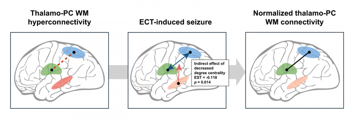
In schizophrenia spectrum disorder, thalamo-parietal white matter hyperconnectivity decreased after ECT along with improvement in overall symptom severity, an effect linked to reduced centrality of the entorhinal cortex (Kim et al., Brain Stimul, 2025).
Prediction of Course and Treatment Response
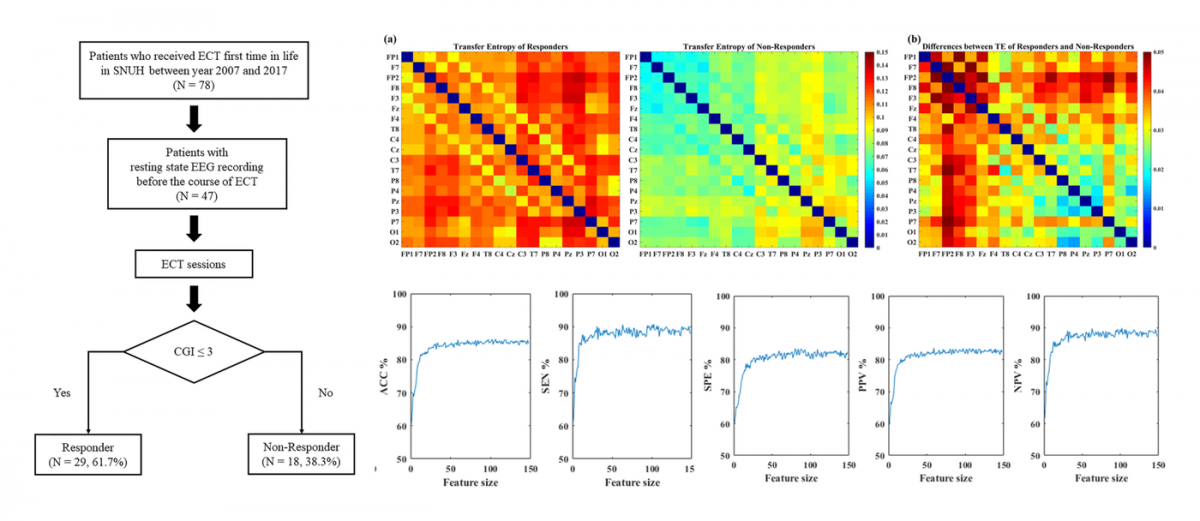
Machine learning analysis of resting-state EEG predicted individual responses to ECT in schizophrenia, with higher frontal effective connectivity linked to a favorable response (Min et al., Schizophr Res, 2020).