


Clinical Cognitive Neuroscience Center
Research
Schizophrenia and High Risk for Psychosis
Schizophrenia and High Risk for Psychosis
Key question. What brain abnormalities underlie schizophrenia, and how do they unfold from clinical high risk to established illness? Building on this, can brain and clinical measures tell us early who will become ill and who will recover?
Approach. We follow individuals at clinical high risk (CHR) and with first-episode psychosis (FEP) in the Seoul Youth Clinic cohort, and study schizophrenia across its course. We combine EEG/ERP, structural, diffusion and functional MRI, and PET with repeated clinical and cognitive assessment. We are a site of the AMP SCZ (ProNET) and PSYSCAN studies and take part in the ENIGMA Schizophrenia and Clinical High Risk working groups.
Recent Findings
Pathophysiological Mechanisms
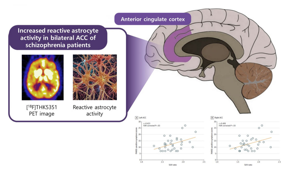
Reactive astrocyte activity, measured with [18F]THK5351 PET, was increased in the bilateral anterior cingulate cortex of patients with schizophrenia and correlated with positive symptom severity (Kim et al., JAMA Netw Open, 2024).
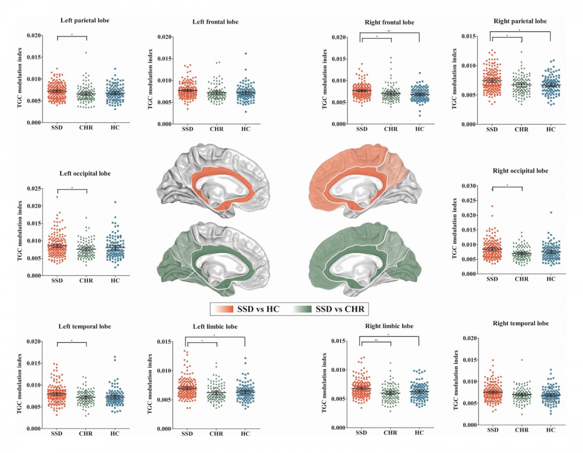
Thalamocortical dysrhythmia was found in patients with schizophrenia spectrum disorder and individuals at CHR (Kim et al., Neuropsychopharmacology, 2022).
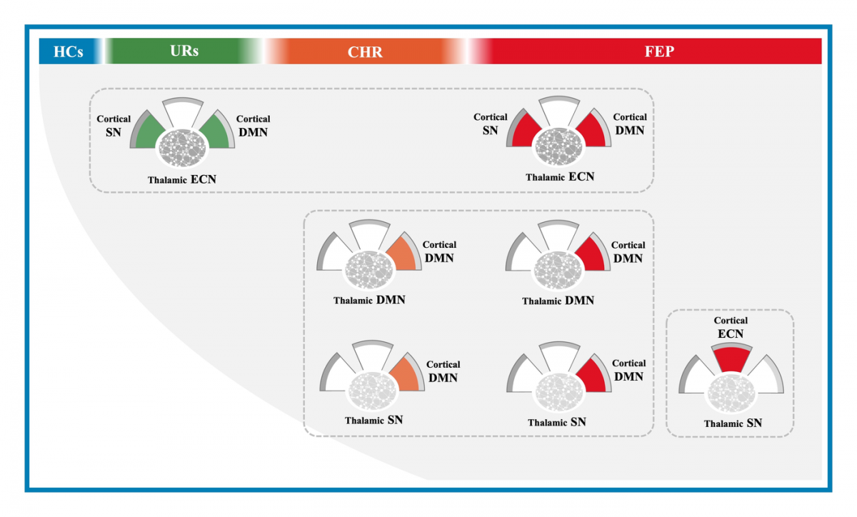
Large-scale thalamocortical triple-network dysconnectivity was present in first-episode psychosis and in individuals at risk for psychosis (Kim et al., Schizophr Bull, 2023).
MRI texture analysis revealed stagewise, nonlinear alterations of frontal gray matter in early psychosis (Moon et al., Mol Psychiatry, 2023).
Prediction of Course and Treatment Response
We developed and internally validated a personalized risk calculator to predict the onset of psychosis in individuals at CHR (Lee et al., Psychol Med, 2022).
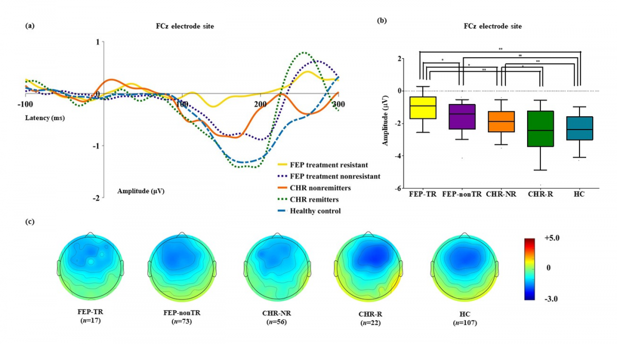
Mismatch negativity measured in early psychosis forecasts prognostic trajectories (Kim et al., Psychol Med, 2023).
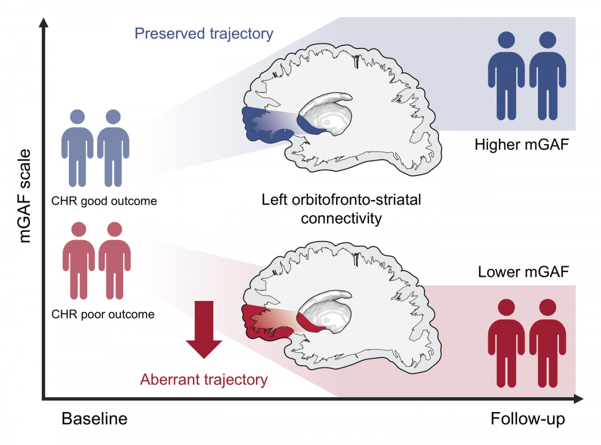
In CHR individuals, corticostriatal structural connectivity follows different trajectories depending on functional outcome (Choe et al., Transl Psychiatry, 2025).
Foundational Work
Mismatch negativity predicted remission in individuals at CHR (Kim et al., Schizophr Bull, 2018).